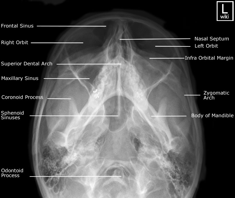
Facial Bones X Ray Om 30 . when the central ray is angled 30 degrees caudad, the petrous ridges are projected below the inferior margins of the orbits. facial bones radiographic anatomy. This case is an example of a normal facial. No displaced facial or skull fracture noted. This page contains radiographic anatomy of the adult facial bones. both the om and om 30 views will identify these fractures, along with the associated soft tissue swelling over the zygomatic. 1 public playlist includes this case. normal appearances of occipitomental (om) 30° view of the skull.
from www.wikiradiography.net
both the om and om 30 views will identify these fractures, along with the associated soft tissue swelling over the zygomatic. when the central ray is angled 30 degrees caudad, the petrous ridges are projected below the inferior margins of the orbits. facial bones radiographic anatomy. This page contains radiographic anatomy of the adult facial bones. This case is an example of a normal facial. normal appearances of occipitomental (om) 30° view of the skull. No displaced facial or skull fracture noted. 1 public playlist includes this case.
Facial Bones Radiographic Anatomy wikiRadiography
Facial Bones X Ray Om 30 when the central ray is angled 30 degrees caudad, the petrous ridges are projected below the inferior margins of the orbits. This case is an example of a normal facial. facial bones radiographic anatomy. 1 public playlist includes this case. both the om and om 30 views will identify these fractures, along with the associated soft tissue swelling over the zygomatic. No displaced facial or skull fracture noted. normal appearances of occipitomental (om) 30° view of the skull. when the central ray is angled 30 degrees caudad, the petrous ridges are projected below the inferior margins of the orbits. This page contains radiographic anatomy of the adult facial bones.
From www.pinterest.co.kr
Radiographic Anatomy of Facial Bones PosteroAnterior Caldwell View Facial Bones X Ray Om 30 facial bones radiographic anatomy. This page contains radiographic anatomy of the adult facial bones. when the central ray is angled 30 degrees caudad, the petrous ridges are projected below the inferior margins of the orbits. both the om and om 30 views will identify these fractures, along with the associated soft tissue swelling over the zygomatic. This. Facial Bones X Ray Om 30.
From radiopaedia.org
Image Facial Bones X Ray Om 30 This case is an example of a normal facial. No displaced facial or skull fracture noted. when the central ray is angled 30 degrees caudad, the petrous ridges are projected below the inferior margins of the orbits. both the om and om 30 views will identify these fractures, along with the associated soft tissue swelling over the zygomatic.. Facial Bones X Ray Om 30.
From dontforgetthebubbles.com
Facial bone xrays Facial Bones X Ray Om 30 normal appearances of occipitomental (om) 30° view of the skull. No displaced facial or skull fracture noted. 1 public playlist includes this case. facial bones radiographic anatomy. when the central ray is angled 30 degrees caudad, the petrous ridges are projected below the inferior margins of the orbits. both the om and om 30 views will. Facial Bones X Ray Om 30.
From www.pinterest.com
Facial Bones Radiographic Anatomy wikiRadiography Medical Facial Bones X Ray Om 30 normal appearances of occipitomental (om) 30° view of the skull. This case is an example of a normal facial. facial bones radiographic anatomy. This page contains radiographic anatomy of the adult facial bones. No displaced facial or skull fracture noted. both the om and om 30 views will identify these fractures, along with the associated soft tissue. Facial Bones X Ray Om 30.
From www.radiologymasterclass.co.uk
Trauma Xray Axial skeleton Face Facial Bones X Ray Om 30 This page contains radiographic anatomy of the adult facial bones. both the om and om 30 views will identify these fractures, along with the associated soft tissue swelling over the zygomatic. when the central ray is angled 30 degrees caudad, the petrous ridges are projected below the inferior margins of the orbits. 1 public playlist includes this case.. Facial Bones X Ray Om 30.
From www.radiologymasterclass.co.uk
Trauma Xray Axial skeleton gallery 1 Facial bones Normal anatomy Facial Bones X Ray Om 30 when the central ray is angled 30 degrees caudad, the petrous ridges are projected below the inferior margins of the orbits. This case is an example of a normal facial. facial bones radiographic anatomy. both the om and om 30 views will identify these fractures, along with the associated soft tissue swelling over the zygomatic. No displaced. Facial Bones X Ray Om 30.
From www.rxdentistry.net
RxDentistry Radiographic Anatomy of Facial Bones Facial Bones X Ray Om 30 This case is an example of a normal facial. facial bones radiographic anatomy. both the om and om 30 views will identify these fractures, along with the associated soft tissue swelling over the zygomatic. No displaced facial or skull fracture noted. normal appearances of occipitomental (om) 30° view of the skull. when the central ray is. Facial Bones X Ray Om 30.
From www.imageinterpretation.co.uk
The Facial Bones Facial Bones X Ray Om 30 both the om and om 30 views will identify these fractures, along with the associated soft tissue swelling over the zygomatic. This case is an example of a normal facial. This page contains radiographic anatomy of the adult facial bones. 1 public playlist includes this case. normal appearances of occipitomental (om) 30° view of the skull. when. Facial Bones X Ray Om 30.
From www.grepmed.com
Facial Bone XRay Anatomy AP by Dr. Naveen Sharma GrepMed Facial Bones X Ray Om 30 both the om and om 30 views will identify these fractures, along with the associated soft tissue swelling over the zygomatic. No displaced facial or skull fracture noted. normal appearances of occipitomental (om) 30° view of the skull. This case is an example of a normal facial. 1 public playlist includes this case. This page contains radiographic anatomy. Facial Bones X Ray Om 30.
From www.pinterest.co.uk
The Facial Bones Facial bones, Maxillary sinus, Herniated Facial Bones X Ray Om 30 This page contains radiographic anatomy of the adult facial bones. when the central ray is angled 30 degrees caudad, the petrous ridges are projected below the inferior margins of the orbits. 1 public playlist includes this case. normal appearances of occipitomental (om) 30° view of the skull. This case is an example of a normal facial. both. Facial Bones X Ray Om 30.
From www.pinterest.es
Radiographic Anatomy PA Caldwell Facial Bones Radiology, Facial Facial Bones X Ray Om 30 This page contains radiographic anatomy of the adult facial bones. No displaced facial or skull fracture noted. when the central ray is angled 30 degrees caudad, the petrous ridges are projected below the inferior margins of the orbits. This case is an example of a normal facial. both the om and om 30 views will identify these fractures,. Facial Bones X Ray Om 30.
From dentallecnotes.blogspot.com
Dentistry lectures for MFDS/MJDF/NBDE/ORE Radiographic Anatomy of Facial Bones X Ray Om 30 This case is an example of a normal facial. facial bones radiographic anatomy. normal appearances of occipitomental (om) 30° view of the skull. No displaced facial or skull fracture noted. 1 public playlist includes this case. when the central ray is angled 30 degrees caudad, the petrous ridges are projected below the inferior margins of the orbits.. Facial Bones X Ray Om 30.
From www.imageinterpretation.co.uk
The Facial Bones Facial Bones X Ray Om 30 facial bones radiographic anatomy. when the central ray is angled 30 degrees caudad, the petrous ridges are projected below the inferior margins of the orbits. normal appearances of occipitomental (om) 30° view of the skull. No displaced facial or skull fracture noted. This page contains radiographic anatomy of the adult facial bones. This case is an example. Facial Bones X Ray Om 30.
From www.wikiradiography.net
Facial Bone Radiography wikiRadiography Facial Bones X Ray Om 30 This page contains radiographic anatomy of the adult facial bones. both the om and om 30 views will identify these fractures, along with the associated soft tissue swelling over the zygomatic. This case is an example of a normal facial. normal appearances of occipitomental (om) 30° view of the skull. facial bones radiographic anatomy. No displaced facial. Facial Bones X Ray Om 30.
From www.wikiradiography.net
Nasal Bones Radiographic Anatomy wikiRadiography Facial Bones X Ray Om 30 1 public playlist includes this case. This page contains radiographic anatomy of the adult facial bones. No displaced facial or skull fracture noted. facial bones radiographic anatomy. when the central ray is angled 30 degrees caudad, the petrous ridges are projected below the inferior margins of the orbits. This case is an example of a normal facial. . Facial Bones X Ray Om 30.
From radtexts.blogspot.com
PA AXIAL PROJECTION FACIAL BONES RadTechOnDuty Facial Bones X Ray Om 30 This case is an example of a normal facial. No displaced facial or skull fracture noted. when the central ray is angled 30 degrees caudad, the petrous ridges are projected below the inferior margins of the orbits. facial bones radiographic anatomy. normal appearances of occipitomental (om) 30° view of the skull. both the om and om. Facial Bones X Ray Om 30.
From fity.club
X Ray Skull Views Facial Bones X Ray Om 30 when the central ray is angled 30 degrees caudad, the petrous ridges are projected below the inferior margins of the orbits. facial bones radiographic anatomy. both the om and om 30 views will identify these fractures, along with the associated soft tissue swelling over the zygomatic. normal appearances of occipitomental (om) 30° view of the skull.. Facial Bones X Ray Om 30.
From www.pinterest.com
Pin on mrt Facial Bones X Ray Om 30 facial bones radiographic anatomy. No displaced facial or skull fracture noted. This page contains radiographic anatomy of the adult facial bones. This case is an example of a normal facial. normal appearances of occipitomental (om) 30° view of the skull. when the central ray is angled 30 degrees caudad, the petrous ridges are projected below the inferior. Facial Bones X Ray Om 30.